
4
質的画像診断
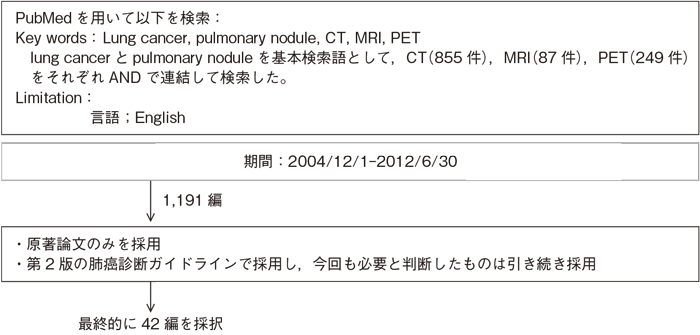
文献検索と採択
4-1.質的画像診断
- 推 奨
- a.高分解能CT(薄層CT)は病理像に対応した特徴的な所見がみられ,高分解能CTを加えることで肺腫瘤性病変の良悪性鑑別に有用な情報を得られる場合があり,行うよう勧められる。(グレードB)
- b.造影CTは,ある程度の除外診断ないし重要な所見の拾い上げが可能で,肺腫瘤性病変の良悪性鑑別の補助診断として,科学的根拠は十分ではないが,行うことを考慮してもよい。(グレードC1)
- c.FDG-PETは,ある程度の除外診断ないし重要な所見の拾い上げが可能で,肺腫瘤性病変の良悪性鑑別の補助診断として,科学的根拠は十分ではないが,行うことを考慮してもよい。(グレードC1)
- d.画像の経時的比較を行うことには慎重な対応が求められる。限定的であるが経時的比較によりある程度の除外診断ないし重要な所見の拾い上げが可能な場合があり,肺腫瘤性病変の良悪性鑑別の補助診断として,科学的根拠は十分ではないが,行うことを考慮してもよい。(グレードC1)
エビデンス
- a.高分解能CTと病理像の対応を検討した多くの研究1)〜7)によって,CT所見の病理学的裏付けが実証され,さらに肺癌の種々の組織型にみられる頻度の高いCT所見が明らかになっている。しかし,良性結節と肺癌の間に所見のオーバーラップがかなりみられること,研究に含まれる結節の大きさや種類によって感度や特異度の値が大きくばらつくことなどの理由で,高分解能CTの肺癌診断能に関する確立したデータはない(感度53〜91%,特異度56〜92%)8)〜10)。孤立性陰影の鑑別診断アルゴリズムには,薄層高分解能CTが使われている11)。CT発見の小型肺野結節の良悪性鑑別診断に高分解能CT画像の使用を勧める12)。また,CAD(computer-aided diagnosis)を用いて,体積計算や造影効果を調べることで診断能の向上を図る試みが報告されている13)14)。
- b.悪性結節は高い造影効果をもつという仮説のもとに,非石灰化の肺野結節の良性悪性の鑑別をヨード造影剤投与後のCTにおける造影効果から判定する方法がある。多施設研究では,15HU以上の造影効果を悪性とした場合の感度は98%,特異度は58%という結果が得られた15)。したがって,造影CTで造影効果がほとんどみられない場合(15HU以下)には強く良性が示唆されるが,造影された場合の質的診断は困難と結論付けられる。
多時相撮像(dynamic study)を行う報告では16)〜18),良性結節を除外診断できる可能性が指摘されている。造影MRIについては,小型肺野孤立陰影の良悪性鑑別や精査必要性の判断に有用であるとする報告がある19)。またDynamic CT,MRI,FDG PET,(99m)Tc-SPECTの間では,良悪性の鑑別能に有意差がないとする報告もある20)。 - c.肺野結節の良性悪性をグルコース代謝の多寡によって判定しようとするFDG-PETは,多くの研究21)〜25)によって,CTより高い診断能をもつことが示された。ただし,定量評価として用いられているstandardized uptake value(SUV)に関しては,比較定量性に問題があるとする報告も多いので,日常診療で標準的な指標として勧められない26)。FDG-PETは悪性病変の診断に高い感度(96.8%)と中等度の特異度(77.8%)をもつことが報告されている27)。ただ,1cm以下の結節のデータは少なく,診断能が確立していないこと,カルチノイド腫瘍や肺胞上皮癌が偽陰性になる場合が多く,肉芽腫の一部は逆に偽陽性になる点に注意が必要である。上記の内容を考慮のうえで,肺野結節の良悪性鑑別診断にFDG-PETを用いることは補助診断として有用である21)〜28)。PET29)〜31)の多時相でSUVを計測する方法(dual PET)の報告がなされ,良性結節を除外診断できる可能性が指摘されている。
PET/CTで良悪性の鑑別を行い良好な成績が報告されている32)33)。FDG-PETなどの機能画像単独よりも,FDG-PET/CTなどの機能画像と形態画像の融合画像が,結節の良悪性鑑別能が高い34)。PET/CTはdynamic CTより診断能が高く,良悪性診断の第一選択はPET/CTと推奨されており,PET/CTが利用できない場合にdynamic CTを代替modaityとするのが合理的とされている35)。 - d.CT技術の進歩によって,小結節が数多く検出されるようになり,その扱いをどのようにすべきかが大きな問題になっている36)37)。CTによる3次元的形態のフォローアップとともに,増大を早期に捉えるために容量測定などの定量的手法が試みられている38)〜41)。また経時的な腫瘍体積計測とPETを併せた所見を用いて良悪性鑑別を行う報告もある42)。
引用文献
- 1)Kodama K, Higashiyama M, Yokouchi H,et al. Prognostic value of ground-glass opacity found in small lung adenocarcinoma on high-resolution CT scanning. Lung Cancer. 2001; 33(1): 17-25. (IV)
- 2)Yang ZG, Sone S, Takashima S, et al. High-resolution CT analysis of small peripheral lung adenocarcinomas revealed on screening helical CT. AJR Am J Roentgenol. 2001; 176(6): 1399-407. (IV)
- 3)Aoki T, Nakata H, Watanabe H, et al. Evolution of peripheral lung adenocarcinomas: CT findings correlated with histology and tumor doubling time. AJR Am J Roentgenol. 2000; 174(3): 763-8. (IV)
- 4)Yabuuchi H, Murayama S, Murakami J, et al. High-resolution CT characteristics of poorly differentiated adenocarcinoma of the peripheral lung: comparison with well differentiated adenocarcinoma. Radiat Med. 2000; 18(6): 343-7. (IV)
- 5)Choi JA, Kim JH, Hong KT, et al. CT bronchus sign in malignant solitary pulmonary lesions: value in the prediction of cell type. Eur Radiol. 2000; 10(8): 1304-9. (IV)
- 6)Furuya K, Murayama S, Soeda H, et al. New classification of small pulmonary nodules by margin characteristics on high-resolution CT. Acta Radiol. 1999; 40(5): 496-504. (IV)
- 7)Koizumi N, Akita S, Sakai K, et al. Classification of air density areas in CT-pathologic correlation of pulmonary adenocarcinoma. Radiat Med. 1995; 13(6): 279-84. (IV)
- 8)Zwirewich CV, Vedal S, Miller RR, et al. Solitary pulmonary nodule: high-resolution CT and radiologic-pathologic correlation. Radiology. 1991; 179(2): 469-76. (IV)
- 9)Seemann MD, Staebler A, Beinert T, et al. Usefulness of morphological characteristics for the differentiation of benign from malignant solitary pulmonary lesions using HRCT. Eur Radiol. 1999; 9(3): 409-17. (IV)
- 10)Takanashi N, Nobe Y, Asoh H, et al. The diagnostic accuracy of a solitary pulmonary nodule, using thin-section high resolution CT: a solitary pulmonary nodule by HRCT. Lung Cancer. 1995; 13(2): 105-12. (IV)
- 11)Libby DM, Smith JP, Altorki NK, et al. Managing the small pulmonary nodule discovered by CT. Chest. 2004 ; 125(4): 1522-9. (IV)
- 12)Li F, Sone S, Abe H, et al. Malignant versus benign nodules at CT screening for lung cancer: comparison of thin-section CT findings. Radiology. 2004; 233(3): 793-8. (IV)
- 13)Awai K, Murao K, Ozawa A, et al. Pulmonary nodules: estimation of malignancy at thin-section helical CT--effect of computer-aided diagnosis on performance of radiologists. Radiology. 2006; 239(1): 276-84. (IV)
- 14)Shah SK, McNitt-Gray MF, Rogers SR, et al. Computer aided characterization of the solitary pulmonary nodule using volumetric and contrast enhancement features. Acad Radiol. 2005; 12(10): 1310-9. (IV)
- 15)Swensen SJ, Viggiano RW, Midthun DE, et al. Lung nodule enhancement at CT: multicenter study. Radiology. 2000; 214(1): 73-80. (IV)
- 16)Jeong YJ, Lee KS, Jeong SY, et al. Solitary pulmonary nodule: characterization with combined wash-in and washout features at dynamic multi-detector row CT. Radiology. 2005; 237(2): 675-83. (IV)
- 17)Bayraktaroglu S, Savas R, Basoglu OK, et al. Dynamic computed tomography in solitary pulmonary nodules. J Comput Assist Tomogr. 2008; 32(2): 222-7. (IV)
- 18)Li Y, Yang ZG, Chen TW, et al. First-pass perfusion imaging of solitary pulmonary nodules with 64-detector row CT: comparison of perfusion parameters of malignant and benign lesions. Br J Radiol. 2010; 83(993): 785-90. (IV)
- 19)Schaefer JF, Vollmar J, Schick F, et al. Solitary pulmonary nodules: dynamic contrast-enhanced MR imaging--perfusion differences in malignant and benign lesions. Radiology. 2004; 232(2): 544-53. (IV)
- 20)Cronin P, Dwamena BA, Kelly AM, et al. Solitary pulmonary nodules: meta-analytic comparison of cross-sectional imaging modalities for diagnosis of malignancy. Radiology. 2008; 246(3): 772-82. (IV)
- 21)Präuer HW, Weber WA, Römer W, et al. Controlled prospective study of positron emission tomography using the glucose analogue [18f]fluorodeoxyglucose in the evaluation of pulmonary nodules. Br J Surg. 1998; 85(11): 1506-11. (IV)
- 22)Lowe VJ, Duhaylongsod FG, Patz EF, et al. Pulmonary abnormalities and PET data analysis: a retrospective study. Radiology. 1997; 202(2): 435-9. (IV)
- 23)Duhaylongsod FG, Lowe VJ, Patz EF Jr, et al. Detection of primary and recurrent lung cancer by means of F-18 fluorodeoxyglucose positron emission tomography (FDG PET). J Thorac Cardiovasc Surg. 1995; 110(1): 130-9. (IV)
- 24)Gupta NC, Maloof J, Gunel E. Probability of malignancy in solitary pulmonary nodules using fluorine-18-FDG and PET. J Nucl Med. 1996; 37(6): 943-8. (IV)
- 25)Gould MK, Maclean CC, Kuschner WG, et al. Accuracy of positron emission tomography for diagnosis of pulmonary nodules and mass lesions: a meta-analysis. JAMA 2001;285:914-24. (IV)
- 26)Detterbeck FC, Falen S, Rivera MP. Seeking a home for a PET, part 1: Defining the appropriate place for positron emission tomography imaging in the diagnosis of pulmonary nodules or masses. Chest. 2004; 125(6): 2294-9. (IV)
- 27)Gould MK, Sanders GD, Barnett PG, et al. Cost-effectiveness of alternative management strategies for patients with solitary pulmonary nodules. Ann Intern Med. 2003; 138(9): 724-35. (IV)
- 28)Lowe VJ, Fletcher JW, Gobar L, et al. Prospective investigation of positron emission tomography in lung nodules. J Clin Oncol. 1998; 16(3): 1075-84. (IV)
- 29)Alkhawaldeh K, Bural G, Kumar R, et al. Impact of dual-time-point (18)F-FDG PET imaging and partial volume correction in the assessment of solitary pulmonary nodules. Eur J Nucl Med Mol Imaging. 2008; 35(2): 246-52. (IV)
- 30)Xiu Y, Bhutani C, Dhurairaj T, et al. Dual-time point FDG PET imaging in the evaluation of pulmonary nodules with minimally increased metabolic activity. Clin Nucl Med. 2007; 32(2): 101-5. (IV)
- 31)Chen CJ, Lee BF, Yao WJ, et al. Dual-phase 18F-FDG PET in the diagnosis of pulmonary nodules with an initial standard uptake value less than 2.5. AJR Am J Roentgenol. 2008; 191(2): 475-9. (IV)
- 32)Yi CA, Lee KS, Kim BT, et al. Tissue characterization of solitary pulmonary nodule: comparative study between helical dynamic CT and integrated PET/CT. J Nucl Med. 2006; 47(3): 443-50. (IV)
- 33)Degirmenci B, Wilson D, Laymon CM, et al. Standardized uptake value-based evaluations of solitary pulmonary nodules using F-18 fluorodeoxyglucose-PET/computed tomography. Nucl Med Commun. 2008; 29(7): 614-22. (IV)
- 34)Tian J, Yang X, Yu L, et al. A multicenter clinical trial on the diagnostic value of dual-tracer PET/CT in pulmonary lesions using 3'-deoxy-3'-18F-fluorothymidine and 18F-FDG. J Nucl Med. 2008; 49(2): 186-94. (IV)
- 35)Yi CA, Lee KS, Kim BT, Choi JY, et al. Tissue characterization of solitary pulmonary nodule: comparative study between helical dynamic CT and integrated PET/CT. J Nucl Med. 2006;47:443-50. (IV)
- 36)Winer-Muram HT. The solitary pulmonary nodule. Radiology. 2006; 239(1): 34-49. (IV)
- 37)MacMahon H, Austin JH, Gamsu G, et al. Guidelines for management of small pulmonary nodules detected on CT scans: a statement from the Fleischner Society. Radiology. 2005; 237(2): 395-400. (IV)
- 38)Yankelevitz DF, Reeves AP, Kostis WJ, et al. Small pulmonary nodules: volumetrically determined growth rates based on CT evaluation. Radiology. 2000; 217(1): 251-6. (IV)
- 39)Hasegawa M, Sone S, Takashima S, et al. Growth rate of small lung cancers detected on mass CT screening. Br J Radiol. 2000; 73(876): 1252-9. (IV)
- 40)Wang JC, Sone S, Feng L, Y, et al. Rapidly growing small peripheral lung cancers detected by screening CT: correlation between radiological appearance and pathological features. Br J Radiol. 2000; 73(873): 930-7. (IV)
- 41)van Klaveren RJ, Oudkerk M, Prokop M, et al. Management of lung nodules detected by volume CT scanning. N Engl J Med. 2009; 361(23): 2221-9. (IV)
- 42)Ashraf H, Dirksen A, Loft A, et al. Combined use of positron emission tomography and volume doubling time in lung cancer screening with low-dose CT scanning. Thorax. 2011; 66(4): 315-9. (IV)
