
4
質的画像診断
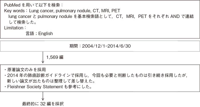
文献検索と採択
4-1.質的画像診断
- 推 奨
- a.高分解能CT(薄層CT)は病理像に対応した特徴的な所見がみられ,高分解能CTを加えることで肺結節の良悪性鑑別に有用な情報を得られるため,行うよう勧められる。(グレードA)
- b.造影CTは,ある程度の除外診断ないし重要な所見が得られ,肺結節の良悪性鑑別の補助診断として行うことを考慮してもよい。(グレードC1)
- c.FDG-PET/CTは,ある程度の除外診断ないし重要な所見が得られ,肺結節の良悪性鑑別の補助診断として行うことを考慮してもよい。(グレードC1)
- d.画像の経過観察を行う際には慎重な対応が求められる。経過観察によりある程度の除外診断ないし重要な所見が得られ,肺結節の良悪性鑑別の補助診断として行うことを考慮してもよい。(グレードC1)
エビデンス
- a.高分解能CTと病理像の対応を検討した多くの研究によって,CT所見の病理学的裏付けが実証され,さらに肺癌の種々の組織型にみられる頻度の高いCT所見が明らかになっている1)~6)。近年MDCTの普及で高分解能CTはCT検査時に簡便に作成できる。孤立性陰影の鑑別診断アルゴリズムには,薄層高分解能CTが使われており7),また,CT発見の小型肺結節の良悪性鑑別診断にも有効である4)。したがって,胸部CTで認められた肺結節に対しては高分解能CTを行うことを強く勧める。ただし,良性結節と肺癌の間に所見のオーバーラップがかなりみられること,研究に含まれる結節の大きさや種類によって感度や特異度の値が大きくばらつくことなどの理由で,高分解能CTの肺癌診断能に関する確立したデータはなく判定の扱いには注意が必要である(感度53~91%,特異度56~92%)9)~11)。
CAD(computer-aided diagnosis)を用いて良悪性鑑別能の向上を図る試み12)なども報告されている。 - b.悪性結節は高い造影効果をもつという仮説のもとに,非石灰化の肺結節の良性悪性の鑑別をヨード造影剤投与後のCTにおける造影効果から判定する方法がある。多施設研究では,15HU以上の造影効果を悪性とした場合の感度は98%,特異度は58%という結果が得られた13)。したがって,造影CTで造影効果がほとんどみられない場合(15HU以下)には強く良性が示唆されるが,造影された場合の質的診断は困難と結論付けられる。
多時相撮像(dynamic study)を行う報告では,良性結節を除外診断できる可能性が指摘されている14)15)。
MRIについては,多時相撮像やDWI(diffusion-weighted magnetic resonance imaging)について小型肺野孤立陰影の良悪性鑑別や精査必要性の判断に有用であるとする報告がある16)17)。またDynamic CT,MRI,FDG PET,(99m)Tc-SPECTの間では,良悪性の鑑別能に有意差がないとする報告もある18)。 - c.肺結節の良性悪性をグルコース代謝の多寡によって判定しようとするFDG-PETは,多くの研究によって,CTより高い診断能をもつことが示唆された19)。ただし,定量評価として用いられているstandardized uptake value(SUV)に関しては,比較定量性に問題があるとする報告も多いので,日常診療で標準的な指標として勧められない20)。また,1cm以下の結節のデータは少なく,診断能が確立していないこと,カルチノイド腫瘍や肺胞上皮癌が偽陰性になる場合が多く,肉芽腫の一部は逆に偽陽性になる点に注意が必要である21)22)。上記の内容を考慮のうえで,肺結節の良悪性鑑別診断にFDG-PETを用いることは補助診断として有用である。
FDG-PET/CTなどの機能画像と形態画像の融合画像が普及し,FDG-PETなどの機能画像単独よりも診断能が高くなることが期待されている。FDG-PET/CTにて良悪性の鑑別を行い良好な成績が報告されている23)24)。多時相でSUVを計測する方法(Dual time point PET/CT)の報告がなされ,良性結節を除外診断できる可能性が指摘されている。 - d.CT技術の進歩によって,小結節が数多く検出されるようになり,その扱いをどのようにすべきかが大きな問題になっている7)26)~28)。現時点での画像診断が困難な肺結節に対し,結節の大きさや性状等の変化を経時的(月単位,年単位)に評価する方法が提案されている29)30)。
臨床情報と画像所見を併せて肺癌の可能性を予測する試み31),CTによる3次元的形態のフォローアップとともに,増大を早期に捉えるために容量測定などの定量的手法が試みられている32),また経時的な腫瘍体積計測とPETを併せた所見を用いて良悪性鑑別を行う報告,などがある33)が測定誤差もあり経過観察には慎重な対応が求められる。
引用文献
- 1)Kodama K, Higashiyama M, Yokouchi H,et al. Prognostic value of ground-glass opacity found in small lung adenocarcinoma on high-resolution CT scanning. Lung Cancer. 2001; 33(1): 17-25. (IV)
- 2)Yang ZG, Sone S, Takashima S, et al. High-resolution CT analysis of small peripheral lung adenocarcinomas revealed on screening helical CT. AJR Am J Roentgenol. 2001; 176(6): 1399-407. (IV)
- 3)Aoki T, Nakata H, Watanabe H, et al. Evolution of peripheral lung adenocarcinomas: CT findings correlated with histology and tumor doubling time. AJR Am J Roentgenol. 2000; 174(3): 763-8. (IV)
- 4)Li F, Sone S, Abe H, et al. Malignant versus benign nodules at CT screening for lung cancer: comparison of thin-section CT findings. Radiology. 2004; 233(3): 793-8. (IV)
- 5)Lee SM, Park CM, Goo JM, et al. Invasive pulmonary adenocarcinomas versus preinvasive lesions appearing as ground-glass nodules: differentiation by using CT features. Radiology. 2013; 268(1): 265-73.(IV)
- 6)Lee KH, Goo JM, Park SJ, et al. Correlation between the size of the solid component on thin-section CT and the invasive component on pathology in small lung adenocarcinomas manifesting as ground-glass nodules. J Thorac Oncol. 2014; 9(1): 74-82.(IV)
- 7)Libby DM, Smith JP, Altorki NK, et al. Managing the small pulmonary nodule discovered by CT. Chest. 2004 ; 125(4): 1522-9. (IV)
- 8)Zwirewich CV, Vedal S, Miller RR, et al. Solitary pulmonary nodule: high-resolution CT and radiologic-pathologic correlation. Radiology. 1991; 179(2): 469-76. (IV)
- 9)Seemann MD, Staebler A, Beinert T, et al. Usefulness of morphological characteristics for the differentiation of benign from malignant solitary pulmonary lesions using HRCT. Eur Radiol. 1999; 9(3): 409-17. (IV)
- 10)Takanashi N, Nobe Y, Asoh H, et al. The diagnostic accuracy of a solitary pulmonary nodule, using thin-section high resolution CT: a solitary pulmonary nodule by HRCT. Lung Cancer. 1995; 13(2): 105-12. (IV)
- 11)Awai K, Murao K, Ozawa A, et al. Pulmonary nodules: estimation of malignancy at thin-section helical CT--effect of computer-aided diagnosis on performance of radiologists. Radiology. 2006; 239(1): 276-84. (IV)
- 12)Swensen SJ, Viggiano RW, Midthun DE, et al. Lung nodule enhancement at CT: multicenter study. Radiology. 2000; 214(1): 73-80. (IV)
- 13)Jeong YJ, Lee KS, Jeong SY, et al. Solitary pulmonary nodule: characterization with combined wash-in and washout features at dynamic multi-detector row CT. Radiology. 2005; 237(2): 675-83. (IV)
- 14)Shan F, Zhang Z, Xing W, et al. Differentiation between malignant and benign solitary pulmonary nodules: use of volume first-pass perfusion and combined with routine computed tomography. Eur J Radiol. 2012; 81(11): 3598-605.(IV)
- 15)Schaefer JF, Vollmar J, Schick F, et al. Solitary pulmonary nodules: dynamic contrast-enhanced MR imaging--perfusion differences in malignant and benign lesions. Radiology. 2004; 232(2): 544-53. (IV)
- 16)Li B, Li Q, Chen C, et al. A systematic review and meta-analysis of the accuracy of diffusion-weighted MRI in the detection of malignant pulmonary nodules and masses. Acad Radiol. 2014; 21(1): 21-9.(IV)
- 17)Cronin P, Dwamena BA, Kelly AM, et al. Solitary pulmonary nodules: meta-analytic comparison of cross-sectional imaging modalities for diagnosis of malignancy. Radiology. 2008; 246(3): 772-82. (IV)
- 18)Gould MK, Maclean CC, Kuschner WG, et al. Accuracy of positron emission tomography for diagnosis of pulmonary nodules and mass lesions: a meta-analysis. JAMA 2001;285:914-24. (IV)
- 19)Gould MK, Sanders GD, Barnett PG, et al. Cost-effectiveness of alternative management strategies for patients with solitary pulmonary nodules. Ann Intern Med. 2003; 138(9): 724-35. (IV)
- 20)Detterbeck FC, Falen S, Rivera MP, et al. Seeking a home for a PET, part 1: Defining the appropriate place for positron emission tomography imaging in the diagnosis of pulmonary nodules or masses. Chest. 2004; 125(6): 2294-9.(IV)
- 21)Erasmus JJ, Macapinlac HA Low-sensitivity FDG-PET studies: less common lung neoplasms. Semin Nucl Med. 2012; 42(4): 255-60.(IV)
- 22)Yi CA, Lee KS, Kim BT, et al. Tissue characterization of solitary pulmonary nodule: comparative study between helical dynamic CT and integrated PET/CT. J Nucl Med. 2006; 47(3): 443-50.(IV)
- 23)Sim YT, Goh YG, Dempsey MF, et al. PET-CT evaluation of solitary pulmonary nodules: correlation with maximum standardized uptake value and pathology. Lung. 2013; 191(6): 625-32.(IV)
- 24)Zhang L, Wang Y, Lei J , et al. Dual time point 18FDG-PET/CT versus single time point 18FDG-PET/CT for the differential diagnosis of pulmonary nodules: a meta-analysis. Acta Radiol. 2013; 54(7): 770-7.(IV)
- 25)Winer-Muram HT. The solitary pulmonary nodule. Radiology. 2006; 239(1): 34-49. (IV)
- 26)van Klaveren RJ, Oudkerk M, Prokop M, et al. Management of lung nodules detected by volume CT scanning. N Engl J Med. 2009; 361(23): 2221-9. (IV)
- 27)de Hoop B, van Ginneken B, Gietema H , et al. Pulmonary perifissural nodules on CT scans: rapid growth is not a predictor of malignancy. Radiology. 2012; 265(2): 611-6.(IV)
- 28)Naidich DP, Bankier AA, MacMahon H, et al. Recommendations for the management of subsolid pulmonary nodules detected at CT: a statement from the Fleischner Society. Radiology. 2013; 266(1): 304-17 .(IV)
- 29)MacMahon H, Austin JH, Gamsu G, et al. Guidelines for management of small pulmonary nodules detected on CT scans: a statement from the Fleischner Society. Radiology. 2005; 237(2): 395-400. (IV)
- 30)McWilliams A, Tammemagi MC, Mayo JR, et al. Probability of cancer in pulmonary nodules detected on first screening CT. N Engl J Med. 2013; 369(10): 910-9.(IV)
- 31)Yankelevitz DF, Reeves AP, Kostis WJ, et al. Small pulmonary nodules: volumetrically determined growth rates based on CT evaluation. Radiology. 2000; 217(1): 251-6. (IV)
- 32)Ashraf H, Dirksen A, Loft A, et al. Combined use of positron emission tomography and volume doubling time in lung cancer screening with low-dose CT scanning. Thorax. 2011; 66(4): 315-9. (IV)
