
2
Ⅱ.非小細胞肺癌(NSCLC)
放射線治療基本的事項
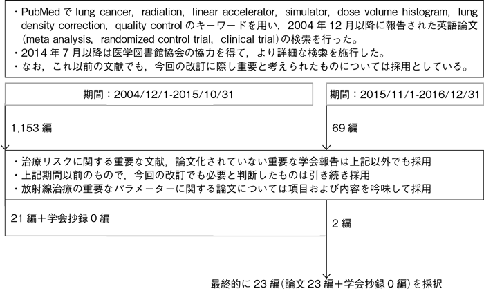
文献検索と採択
本文中に用いた略語および用語の解説
| DVH | dose-volume histogram | 線量体積ヒストグラム |
|---|---|---|
| MLD | mean lung dose | 平均肺線量 |
| V20 | 20 Gy以上照射される肺体積の全肺体積に対する割合 | |
| V30 | 30 Gy以上照射される肺体積の全肺体積に対する割合 | |
2-1.放射線治療装置・治療計画法
- 推 奨
a.肺癌に対する胸部放射線治療には直線加速器による6~10 MV X線を用いるよう勧められる。
b.放射線治療計画には,CTシミュレーションによる3次元治療計画を行うよう勧められる。
c.肺癌の放射線治療では,できるかぎり実測値に近い計算アルゴリズムを用いた不均質肺補正を行い,3次元的な線量分布を常に検討することを行うよう勧められる。
エビデンス
- a.
- 肺癌の胸部放射線治療では直線加速器による高エネルギーX線が用いられるが,エネルギーが低いと照射範囲内の線量不均一性が高度となり,逆にエネルギーが高すぎても標的辺縁ではビルドアップ効果により線量の低下を招く1)~4)。このようにX線の物理的特性から至適エネルギーとして6~10 MV X線の使用が推奨される。ただし,定位放射線照射の場合には4~6 MV X線が望ましい。
- b.
- 3次元治療計画により,ターゲットの線量を低下させることなく正常肺と心臓の平均線量を有意に減少できることが示されている5)~7)。また,放射線治療単独例に対し,肺のDVHと放射線肺臓炎の関係について検討され,Grade 2(RTOGの基準)以上の放射線肺臓炎の発症リスクを低下させるには,V20が40%を超えないようにすることが重要であると報告されている8)。また,化学療法併用の際には,V20が25%を超えないように治療計画することを推奨している9)。さらに,全肺のV20だけではなくV30やMLDなどのパラメーターと放射線肺臓炎の発生との相関についても報告されている10)~12)。その他,放射線食道炎と関連する食道のパラメーター13)14), 心毒性と相関する心臓のパラメーター15)などについても報告され,リスク評価における有用性が示唆されており,放射線治療計画には,CTシミュレーションによる3次元治療計画を行うよう勧められる。
- c.
- ファントムを用いた線量測定実験で,肺内孤立性腫瘍を10 MV X線で照射した場合,肺補正なしでは,線量は10~20%の過線量となる。一方,肺補正を行うと線量計算アルゴリズムによって8~18%の線量不足となる16)。臨床の肺癌症例での検討では,肺補正を行わないと5~28%の過線量となると報告されている16)~18)。また,肺野型腫瘍に対してはエネルギーの低いX線を用いたほうが良好な線量分布を得られると報告されている19)。計算アルゴリズム精度も向上しており,実地医療において3次元的な線量分布を検討するよう勧められる。
2-2.放射線療法の品質管理
- 推 奨
- 放射線療法では,照射野設定,線量計算などの品質管理を適切に行うよう勧められる。
エビデンス
引用文献
- 1)Phlips P, Rocmans P, Vanderhoeft P, et al. Postoperative radiotherapy after pneumonectomy: impact of modern treatment facilities. Int J Radiat Oncol Biol Phys. 1993; 27(3): 525-9.
- 2)DesRosiers PM, Moskvin VP, DesRosiers CM, et al. Lung cancer radiation therapy: Monte Carlo investigation of“under dose”by high energy photons. Technol Cancer Res Treat. 2004; 3(3): 289-94.
- 3)Osei EK, Darko J, Mosseri A, el al. EGSNRC Monte Carlo study of the effect of photon energy and field margin in phantoms simulating small lung lesions. Med Phys. 2003; 30(10): 2706-14.
- 4)Wang L, Yorke E, Desobry G, et al. Dosimetric advantage of using 6 MV over 15 MV photons in conformal therapy of lung cancer: Monte Carlo studies in patient geometries. J Appl Clin Med Phys. 2002; 3(1): 51-9.
- 5)Ragazzi G, Cattaneo GM, Fiorino C, et al. Use of dose-volume histograms and biophysical models to compare 2D and 3D irradiation techniques for non-small cell lung cancer. Br J Radiol. 1999; 72(855): 279-88.
- 6)Schraube P, von Kampen M, Oetzel D, et al. The impact of 3-D radiotherapy planning after a pneumonectomy compared to a conventional treatment set-up. Radiother Oncol. 1995; 37(1): 65-70.
- 7)DiBiase SJ, Werner-Wasik M, Croce R, et al. Standard off-cord lung oblique fields do not include the entire mediastinum: a computed tomography simulator study. Am J Clin Oncol. 2000; 23(3): 249-52.
- 8)Graham MV, Purdy JA, Emami B, et al. Clinical dose-volume histogram analysis for pneumonitis after 3D treatment for non-small cell lung cancer(NSCLC). Int J Radiat Oncol Biol Phys. 1999; 45(2): 323-9.
- 9)Tsujino K, Hirota S, Endo M, et al. Predictive value of dose-volume histogram parameters for predicting radiation pneumonitis after concurrent chemoradiation for lung cancer. Int J Radiat Oncol Biol Phys. 2003; 55(1): 110-5.
- 10)Hernando ML, Marks LB, Bentel GC, et al. Radiation-induced pulmonary toxicity: a dose-volume histogram analysis in 201 patients with lung cancer. Int J Radiat Oncol Biol Phys. 2001; 51(3): 650-9.
- 11)Fay M, Tan A, Fisher R, et al. Dose-volume histogram analysis as predictor of radiation pneumonitis in primary lung cancer patients treated with radiotherapy. Int J Radiat Oncol Biol Phys. 2005; 61(5): 1355-63.
- 12)Ramella S, Trodella L, Mineo TC, et al. Adding ipsilateral V20 and V30 to conventional dosimetric constraints predicts radiation pneumonitis in stage IIIA-B NSCLC treated with combined-modality therapy. Int J Radiat Oncol Biol Phys. 2010; 76(1): 110-5.
- 13)Rose J, Rodrigues G, Yaremko B, et al. Systematic review of dose-volume parameters in the prediction of esophagitis in thoracic radiotherapy. Radiother Oncol. 2009; 91(3): 282-7.
- 14)Pan Y, Brink C, Knap M, et al. Acute esophagitis for patients with local-regional advanced non small cell lung cancer treated with concurrent chemoradiotherapy. Radiother Oncol. 2016; 118(3): 465-70.
- 15)Ming X, Feng Y, Yang C, et al. Radiation-induced heart disease in lung cancer radiotherapy: A dosimetric update. Medicine (Baltimore). 2016; 95(41): e5051.
- 16)Yorke E, Harisiadis L, Wessels B, et al. Dosimetric considerations in radiation therapy of coin lesions of the lung. Int J Radiat Oncol Biol Phys. 1996; 34(2): 481-7.
- 17)Orton CG, Chungbin S, Klein EE, et al. Study of lung density corrections in a clinical trial(RTOG 88-08). Radiation Therapy Oncology Group. Int J Radiat Oncol Biol Phys. 1998; 41(4): 787-94.
- 18)van’t Riet A, Stam HC, Mak AC, et al. Implications of lung corrections for dose specification in radiotherapy. Int J Radiat Oncol Biol Phys. 1985; 11(3): 621-5.
- 19)Klein EE, Morrison A, Purdy JA, et al. A volumetric study of measurements and calculations of lung density corrections for 6 and 18 MV photons. Int J Radiat Oncol Biol Phys. 1997; 37(5): 1163-70.
- 20)White JE, Chen T, McCracken J, et al. The influence of radiation therapy quality control on survival, response and sites of relapse in oat cell carcinoma of the lung: preliminary report of a Southwest Oncology Group study. Cancer. 1982; 50(6): 1084-90.
- 21)Schaake-Koning C, Kirkpatrick A, Kröger R, et al. The need for immediate monitoring of treatment parameters and uniform assessment of patient data in clinical trials. A quality control study of the EORTC Radiotherapy and Lung Cancer Cooperative Groups. Eur J Cancer. 1991; 27(5): 615-9.
- 22)Wallner PE, Lustig RA, Pajak TF, et al. Impact of initial quality control review on study outcome in lung and head/neck cancer studies--review of the Radiation Therapy Oncology Group experience. Int J Radiat Oncol Biol Phys. 1989; 17(4): 893-900.
- 23)Groom N, Wilson E, Lyn E, et al. Is pre-trial quality assurance necessary? Experiences of the CONVERT Phase III randomized trial for good performance status patients with limited-stage small-cell lung cancer. Br J Radiol. 2014; 87(1037): 20130653.
